PORTFOLIO
CROSSBRAIN was funded as part of a portfolio of projects under the same call. We place great value on cross project collaboration with our sister projects and will dedicate significant amount of resources to on portfolio activities, i.e. cross-project collaboration.
CITRUS – Closed-loop Individualized image-guided Transcranial Ultrasonic Stimulation
Grant agreement ID: 101071008
Objective: We are joining forces across Europe to advance a new non-invasive technology – transcranial ultrasound stimulation (TUS) – to reversibly modulate brain regions with exquisite millimetre precision, even deep in the brain. As such, we aim to establish an urgently needed novel treatment option for neurological and psychiatric diseases. TUS combines the precision and reach of invasive deep brain stimulation, required to directly target clinically relevant structures, with the non-invasive and low-cost nature of transcranial electromagnetic techniques that are inherently limited in focus and depth. The main roadblock to widespread adoption of TUS in neuroscientific and clinical applications is the difficulty of steering the small ultrasound focus onto the intended target and reaching the desired intensity, with no empirical validation of targeting success currently available. We will develop a neuronavigated TUS-MRI system with advanced magnetic resonance imaging (MRI)-guided application planning and closed-loop application control to enable safe, individualised, and effective high-precision TUS in humans. As such we will unlock the full potential of TUS to non-invasively modulate deep brain structures with unprecedented spatial precision in the millimetre range. The final prototype will be a fully functional device that integrates novel MR-compatible 256-element TUS-transducers (for advanced 3D-steering of the TUS focus) with a custom-tailored 32-channel MR-receiver coil (for accelerated imaging with maximal sensitivity) and closed-loop target validation using MR-acoustic radiation force imaging (MR-ARFI). This novel device with its unique features will enable for the first time the personalized non-invasive high-precision stimulation of cortical and subcortical targets in the human brain. It will be a game changer for both neuroscientific research and clinical application in neurological and psychiatric diseases with the potential to benefit millions of patients.
Citrus
Hyperstim
HYPERSTIM- High-dimensional electrical stimulation for visual prosthesis
Grant agreement ID: 101071015
Project description: Electrodes to restore vision: Future advanced neuroprostheses, especially visual prostheses, will need to transfer much more information than currently possible. Improving the electrode count will be part of the solution. The EU-funded HYPERSTIM project will use the available electrodes more efficiently by applying sophisticated stimulation protocols. The project’s main objective is to achieve a resolution of at least 20 times the number of electrodes that are physically present with a combination of techniques. The obtained order-of-magnitude improvement in resolution will spur the development of breakthrough prostheses that will be widely adopted by blind patients and bring the field of neural interfacing to the next level.
Objective: Future advanced neuroprostheses will need to transfer orders of magnitude more information to the brain than currently possible. This is most urgently needed in visual prostheses. Improving the electrode count will be part of the solution: a next generation of visual prosthesis will most probably be based on the insertion of over 1000 microelectrodes in the visual cortex. Still, current visual prostheses use very simple stimulation patterns, in which at most the stimulation amplitude is modulated. We propose to explore a second, complementary approach to brute scaling: using the available electrodes more efficiently by applying sophisticated stimulation protocols. Our main objective is to achieve a fundamental breakthrough in the spatial resolution of electrical brain stimulation to restore vision, obtaining a resolution of at least 20X the number of electrodes that are physically present. The vast number of possible stimulation combinations calls for a radically new research methodology, integrating modeling and state-of-the-art neuroscience methods at every spatial scale (from single neurons to the entire brain) in a closed-loop optimization process. With this combination of techniques, we will study which stimulation patterns effectively induce sufficient neural activations in higher areas (i.e. ignition) and cause visual perceptions. Thus, we will be able to explore the vast, hyperdimensional search space of possible stimulation patterns, and produce a set of in vivo tested stimulation patterns that are capable of eliciting distinguishable physiological and behavioral responses. The obtained order-of-magnitude improvement in resolution will spur the development of breakthrough prostheses that will be widely adopted by blind patients, and bring the field of neural interfacing to the next level.
HYPERPROBE – Transforming brain surgery by advancing functional-guided neuronavigational imaging
Grant agreement ID: 101071040
Project description: Innovative neuronavigational technology for brain surgery. Neuronavigation enables neurosurgeons to avoid functional brain locations when removing tumours, using computer-assisted technologies. However, the inability to maintain accurate spatial information on lesions intraoperatively remains a challenge for brain tumour surgeries. The EU-funded HyperProbe project will assemble an interdisciplinary team to develop an innovative optical intraoperative imaging system based on hyperspectral imaging (HSI) and artificial intelligence (AI), for image reconstruction and molecular fingerprinting. The novel HSI system will be handheld and user-friendly, applying AI-based algorithms for the analysis and quantification of images. The project aims to validate the developed technology in vivo, using gold standard modalities in neuronavigational imaging, and provide final proof-of-principle during brain tumour surgery.
Objective: In recent years, through the advancement of imaging technologies (such as MRI, PET, CT, among others) clinical localisation of lesions of the central nervous system (CNS) pre-surgery has made possible for neurosurgeons to plan and navigate away from functional brain locations when removing tumours. However, neuronavigation in the surgical management of brain tumours remains a significant challenge, due to the inability to maintain accurate spatial information of lesioned and non-lesioned locations intraoperatively. To answer this challenge, we have put together a team of engineers, physicists, data scientists and neurosurgeons to develop an innovative, all-optical intraoperative imaging system based on (i) hyperspectral imaging (HSI) for rapid, multi wavelength spectral acquisition, and (ii) artificial intelligence (AI) for image reconstruction and molecular fingerprint recognition. Our intraoperative HSI system (HyperProbe) will (1) map, monitor and quantify biomolecules of interest; (2) be handheld and user-friendly; (3) apply AI-based methods for the reconstruction of spectral images, the analysis of spatio-spectral data and the development and quantification of novel biomarkers. We will validate the developed capacity in phantoms, in vivo against gold standard modalities in neuronavigational imaging, and finally provide proof-of principle during brain tumour surgery. HyperProbe aims at providing functional and structural information on biomarkers of interest that is currently missing during neuro-oncological interventions.
Hyperprobe
Microvasc
MICROVASC – Remote whole-brain functional microscopy of the vascular system: a paradigm shift for the monitoring and treatment of small vessel diseases
Grant agreement ID: 101070917
Project description: Innovative whole-brain functional neuroimaging. Understanding of brain neurovascular coupling and haemodynamic/neurovascular abnormalities as well as evaluation of drug efficacy require high-quality non-invasive neuroimaging of a living brain. The EU-funded MICROVASC project aims to revolutionise neuroimaging with innovative functional ultrasound localisation microscopy (FULM) for transcranial microscopic monitoring of the whole brain vasculature and function. FULM will increase the spatial resolution of brain angiography by at least two orders of magnitude and enable mapping of the functional brain response during task-related and spontaneous activity. The project objectives include the development of advanced neurocomputational analytical methods and validation in preclinical models of cerebrovascular diseases, concluding with the first-in-human study.
Objective: Obtaining functional information on living organs non-invasively across different size scales is a tremendous challenge in medical imaging research, as diseases start locally at the cellular level deep into organs before expressing large-scale and observable symptoms. The unique complexity of the human brain adds another level of difficulty for neuroimaging. The cerebrovascular system consists of a multiscale network of blood vessels. Interaction between neurons and this vascular system, the so-called neurovascular coupling, is a major foundation of brain function leading to constant adaptation of the local cerebral blood flow to local metabolic demand. Its alteration is intimately linked to cerebral dysfunction. Current brain imaging modalities are essential for evaluating cerebrovascular diseases in patients but are restricted to millimetric resolution and fail to capture most of blood flow dynamics. Here, we propose to revolutionize the field of neuroimaging by introducing a groundbreaking technology called functional Ultrasound Localization Microscopy (fULM) capable of monitoring transcranially the whole human brain vasculature and function down to microscopic resolution. Beyond opening a complete paradigm shift in brain angiography (at least two orders of magnitude increase in spatial resolution), fULM will also be able to map the functional brain response during task-evoked and spontaneous activity at microscopic levels. We will address major technical challenges of ultrasound imaging, develop advanced neurocomputational analysis methods, validate our methods in preclinical models of cerebrovascular diseases and perform a First-In-Human study. Fundamental understanding of brain hemodynamics and neurovascular coupling as well as early clinical diagnosis of neurovascular abnormalities and evaluation of drug efficacy would tremendously benefit from such capabilities revealing both the brain vasculature and neurofunctional activity down to microscopic resolutions.
NEMO-BMI – Auto-adaptive Neuromorphic Brain Machine Interface: toward fully embedded neuroprosthetics
Grant agreement ID: 101070891
Project description: Nearly 746,000 people sustain a spinal cord injury every year, with dramatic human, societal and economical cost, leading to impairment or even complete loss of motor functions. Motor Brain-Machine Interfaces (BMIs) translate brain neural signals into commands to external effectors. BMIs raise hopes that limb mobility may be restored, providing patients with control over orthoses, prostheses, or over their own limbs using electrical stimulation. In spite of spectacular results, taking neuroprosthetics into daily practice has proven difficult. Currently, neuroprosthetics are restricted to assisted trials in laboratories, and require regular retraining of a decoder in a supervised manner within controlled environments. They include various components (recording device, antennas, base station, computers connected to effectors, etc.) that are complicated to install and to use. Building on the consortium’s unique experience in clinical chronic BMIs, the project will address major methodological and technological breakthroughs to achieve the first assistance-free motor neuroprosthetics system. NEMO BMI project will conduct the exploration of assistance-free and easy to use portable neuroprosthetics including wireless neuronal activity recorder, a real-time neuronal activity decoder based on integrated technologies, and a spinal cord stimulator. A first objective is the crucial improvement of usability, by introducing an auto-adaptive framework to train the decoder in an adaptive manner during the neuroprosthetics unsupervised use. Brain-guided spinal cord stimulation activating patients’ limbs with an automatic stimulus pattern optimization is the second project objective. A third objective is the exploration of miniaturized embedded solutions by taking advantage of a novel neuromorphic hardware architecture. NEMO BMI technologies will be studied offline and online in two ongoing clinical trials, and will be critical to specify the next-generation assistance-free BMI.
Nemo-BMI
Upside
UPSIDE – Focused Ultrasound Personalized Therapy for the Treatment of Depression
Grant agreement ID: 101070931
Project description: Novel stimulation and recording system for treatment-resistant depression patients. Major depressive disorder (MDD) is a mood disorder which can interfere with daily life, and it is a major contributor to the overall global disease burden. About one third of MDD patients do not respond to established treatments. What is more, reliable biomarkers for depression are needed as a diagnostic tool. The EU-funded UPSIDE project aims to address these issues. To that end, it will research and validate in vivo a hybrid neurotechnology consisting of an epidural focused ultrasound stimulator and a high-density epidural EEG recording system. Project work will lead to a demonstrator, paving the way for personalised treatments for treatment-resistant depression patients.
Objective: Major depressive disorder (MDD) is the leading cause of disability worldwide, affecting 300 million people with a lifetime prevalence of 15%. Approximately one third of all MDD patients fail to respond to currently established treatments based on medication and psychotherapy, thus falling into the category of Treatment-Resistant Depression (TRD) patients. Electroconvulsive therapy (ECT), repetitive Transcranial Magnetic Stimulation (tRMS), Vagus nerve stimulation, deep brain stimulation (DBS) and transcranial focused ultrasound (tFUS) still show poor spatial resolution (ECT, tRMS, tFUS) or low network coverage (VNS, DBS), with average remission rates in clinical trials still lower than 30 %. Apart from the existing stimulation hurdles, reliable biomarkers for depression are needed as a diagnostic tool, and, in the case of NT, to determine the stimulation efficacy and allow for personalized treatment. The UPSIDE project proposes a minimally invasive, high spatial resolution and multi-brain region stimulation and recording system to largely exceed the capabilities of existing NT for depression. Our objective is to research and validate in vivo an hybrid neurotechnology consisting of an epidural focused ultrasound (eFUS) stimulator employing three-dimensional beamforming, and a high-density epidural EEG recording system. Epidural deployment of these devices will be enabled by novel methods for massive integration and miniaturization of high-performing piezoelectric ultrasound materials and high-fidelity organic bioelectronic materials with high energy-efficient complementary metal-oxide semiconductor (CMOS) technology in a biocompatible manner. The UPSIDE project will result in a demonstrator which will allow, for the first time, network stimulation and simultaneous biomarker readout in behavioral experiments with animal models featuring depression-like symptoms. This technological breakthrough will pave the way towards a personalized treatment for TRD.
INTRECOM – Intracranial Neuro Telemetry to Restore Communication
Grant agreement ID: 101070939
Project description:
– Unlocking the communication capabilities of patients with locked-in syndrome.
Locked-in syndrome is a rare and serious neurological disorder caused by damage to a part of the brainstem, which leaves a person totally paralysed but conscious and with normal cognitive abilities. This means they can see, hear and feel normally – even a fly on their nose, for example – yet they cannot move or speak. The psychological and emotional burden on patients and their caregivers is tremendous. The EU-funded INTRECOM project is developing a pioneering, fully implantable AI-based brain-computer interface to address this. It will enable real-time decoding of a person’s intended speech, and automatic transmission to receiving equipment in a bid to unlock the potential of these patients to communicate.
Objective: Not being able to communicate while still being conscious is a horrifying prospective for many patients worldwide. Patients with motor neuron disorders, trauma or stroke risk losing complete muscle control leading to Locked-In Syndrome (LIS) which leaves them completely paralysed and unable to communicate. This is an intolerable, fearful situation with very low quality of life and extreme burden of care for patients, family, and care givers. Intracranial Neuro Telemetry to REstore COMmunication (INTRECOM) will provide a breakthrough for these patients by developing a novel, fully implantable Brain-Computer Interface (BCI) technology that allows for real-time speech decoding and use in the home environment. This BCI system will significantly transcend current technology by providing a high-resolution and sustainable device, combining state-of-the-art hardware and software solutions based on Artificial Intelligence (AI) to liberate LIS patients from their isolation.
Intrecom
Portfolio meetings
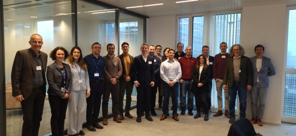
1. Portfolio meeting “Tools to measure and stimulate activity in brain tissue” at EISMEA, Brussels, 21 February 2024
2. Online meeting: Collaborative development and endorsement of a strategic plan and addressing regulatory challenges, 7 October 2024
3. Online meeting: Third Coordination Meeting – BrainTools Portfolio, 28 November 2024
4. Strategic Meeting of the “BrainTools Portfolio” at EISMEA, Brussels, 23-24 April 2025
This project has received funding from the European Union’s European Innovation Council, call HORIZON-EIC-2021-